I've begun to appreciate modern technology in aiding a diagnosis, but not in the way many might think. One of the keys to this case was the video the owner took with her digital camera. Since the pain was sporadic and mainly happened at night, she took the video so a vet could see what was going on. The dog's posture and actions certainly seemed to indicate neck pain. But without symptoms in the clinic, this was puzzling. Well, we knew a problem was in the neck, so we took x-rays of that area.
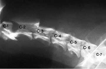
When I looked at the film my breath caught. The problem was immediately obvious and was a serious one. This is an image of the neck of a normal dog.
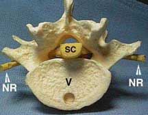
This dog had a severe dislocation of the atlas and axis. Below is an image from the 'Net and not from this case, but my patient was very similar.
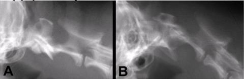
The left image is normal and the right one shows atlantoaxial subluxation. Pay careful attention to the center of the vertebrae where the spinal cord would run. The displacement of the vertebrae bends the spinal cord and puts pressure against it. This can cause pain and was the source of our problems.
The scariest part of this is that this dog is a ticking time bomb. At any time the dog's neck could shift more severely, immediately causing damage to the spinal cord. This can cause paralysis from the neck down, but more importantly can cause problems with the heart and respiratory system since the nerves controlling these vital organs run through the neck. I've seen a couple of cases like this and have always shuddered when I saw the radiographs because of the potential harm that could have happened during the exam. When a patient has neck pain we will manipulate and bend the neck in all directions to see if we can isolate where the problem is. Unfortunately in this case that manipulation can damage the spine.
Thankfully, this is a treatable problem. Surgery can be performed to place pins or plates between the vertebrae once they are realigned, keeping them from moving out of place again. Patients normally do well after this surgery and can lead normal lives afterwards. This dog has an appointment with a surgical specialist and will hopefully have the procedure done soon before this particular bomb goes off.